Periodic eye and vision examinations are an important part of preventive health care. Many eye and vision problems have no obvious signs or symptoms. As a result, individuals are often unaware that problems exist. Early diagnosis and treatment of eye and vision problems are important for maintaining good vision and eye health, and when possible, preventing vision loss.
A complete exam takes 45 minutes on an average. Bring your current glasses with you. If you need a prescription for contact lenses, come to the office wearing contacts. If you are a new patient bring your contact lens details with you. Keep in mind that a dilated exam will preclude reading for 3-4 hours and will cause increased glare for distance.
A comprehensive adult eye and vision examination may include, but is not limited to, the following tests. Individual patient signs and symptoms, along with the professional judgment of the doctor, may significantly influence the testing done.
Patient History
A patient history helps to determine any symptoms the individual is experiencing, when they began, the presence of any general health problems, medications taken and occupational or environmental conditions that may be affecting vision. The doctor will ask about any eye or vision problems you may be having and about your overall health. The doctor will also ask about any previous eye or health conditions of you and your family members.
Visual Acuity

Reading charts are often used to measure visual acuity.
Visual acuity measurements evaluate how clearly each eye is seeing. As part of the testing, you are asked to read letters on distance and near reading charts. The results of visual acuity testing are written as a fraction such as 20/40.
When testing distance vision, the top number in the fraction is the standard distance at which testing is done, twenty feet. The bottom number is the smallest letter size you were able to read. A person with 20/40 visual acuity would have to get within 20 feet of a letter that should be seen at 40 feet in order to see it clearly. Normal distance visual acuity is 20/20.
Preliminary Tests
Preliminary testing may include evaluation of specific aspects of visual function and eye health such as depth perception, color vision, eye muscle movements, peripheral or side vision, and the way your pupils respond to light.
Keratometry
This test measures the curvature of the cornea, the clear outer surface of the eye, by focusing a circle of light on the cornea and measuring its reflection. This measurement is particularly critical in determining the proper fit for contact lenses.
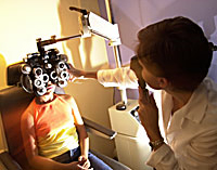
Refraction
Refraction is conducted to determine the appropriate lens power needed to compensate for any refractive error (nearsightedness, farsightedness, or astigmatism). Using an instrument called a phoropter, your optometrist places a series of lenses in front of your eyes and measures how they focus light using a hand held lighted instrument called a retinoscope. The doctor may choose to use an automated instrument that automatically evaluates the focusing power of the eye. The power is then refined by patient’s responses to determine the lenses that allow the clearest vision.
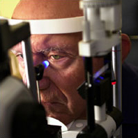
Tonometry
Tonometry measures eye pressure. Elevated pressure in the eye signals an increased risk for glaucoma. Measurement of pressure within the eye (tonometry) is performed. Normal eye pressures range from 10 to 21 millimeters of mercury (mm Hg), averaging about 14 to 16 mm Hg. Anyone with eye pressure greater than 22 mm Hg is at an increased risk of developing glaucoma, although many people with normal pressure also develop glaucoma. Screening tests include puff tonometers and icare which do not use eye drops. But for more accurate testing, numbing drops are used and a special tonometer is used with the help of a blue light to check eye pressure.
External examination of the eye
External examination of the eye includes evaluation of the cornea, eyelids, conjunctiva and surrounding eye tissue using bright light and magnification.
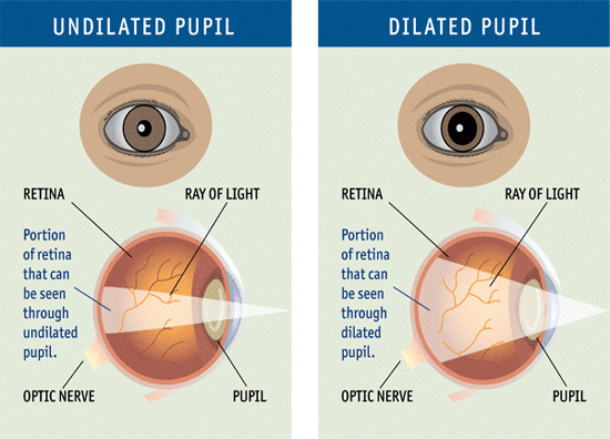
Dilated retinal exam
Evaluation of the lens, retina and posterior section of the eye may be done through a dilated pupil to provide a better view of the internal structures of the eye. Drops are placed in your eyes to dilate, or widen, the pupils. A special magnifying lens to examine your retina to look for signs of damage and other eye problems, such as diabetic retinopathy or age-related macular degeneration. Dilation is the most important part of a comprehensive eye exam because it allows your eye care professional to get a better view of the inside of the eye. However dilation will affect vision for reading and will cause glare if driving for 3-4 hours. While a lot of people can drive out of the office on their own using sunglasses, it may be advisable to bring a driver with you if you feel uncomfortable driving with glare.
Additional testing may be needed based on the results of the previous tests to confirm or rule out possible problems, to clarify uncertain findings, or to provide a more in-depth assessment.
Visual Field Test
This test measures side (peripheral) vision. It helps determine if there is loss of side vision – a sign of glaucoma.
Pachymetry
A numbing drop is applied to the eye. An ultrasound wave instrument is used to measure the thickness of the cornea.
Optical Coherence Tomography or OCT
OCT is a noncontact, noninvasive imaging technique used to obtain high resolution cross-sectional images of the retina. OCT is analogous to ultrasound B-scan imaging except that light rather than sound waves are used in order to obtain a much higher longitudinal resolution of approximately 10µm in the retina. OCT has been shown to be clinically useful for imaging selected macular diseases including macular holes, macular edema, age-related macular degeneration, central serous chorioretinopathy and epiretinal membranes.
In addition, OCT has the capability of measuring the retinal nerve fiber layer thickness in glaucoma and other diseases of the optic nerve.
VEP and ERG
Electrophysiological testing to test optic nerve pathways and retinal function for early detection of glaucoma or retinal disorders.